

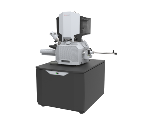
초저온집속이온빔장치 (Cryo-FIB)
| 사진 |
 |
|---|---|
| 영문명/단축명 | Cryo Focused Ion Beam/Scanning Electron Microscope / Cryo-FIB/SEM |
| 모델명 |
Aquilos 2 |
| 설치장소 | 미세구조분석실 / [82113] FIB lab.2 |
| 제작사 | Thermo Fisher |
| 도입년도/가격 | 2023.9월 / 원 |
| 담당자/연락처/e-mail | 박정양(PARK JEONG YANG) / 031-299-6726 / kiki0409@skku.edu |
♦ Features
• 자동화된 batch cryo-lamella milling
- 세포의 특정 관심 지점을 표시하여 여러 개의 라멜라를 자동으로 준비
• Cryo-lift-out 라멜라 준비
- 나노미터 수준의 위치 정확도로 특정 표적 영역의 라멜라 준비 가능
• 3D visualization
- Cryo-Auto Slice and View 소프트웨어를 통해 순차적 milling된 시료 단면에 대한 3차원 이미지 획득
• iFLM Correlative System
- 형광 표시된 세포를 높은 진공 상태에서 이미지화하여 특정 관심 지점을 확인하여 정확하게 시료 준비
♦ Additional Options
• Cryo Sample Preparation with embedded Fluorescence microscope(iFLM) & sputter coater
• Automated Cryo Sample Preparation with AutoTEM for on-the-grid batch lamella
• 3D Volume Imaging with Auto Slice & View software
• Large 2D Imaging Analysis with tiling & stitching
♦ Specification
• Electron Gun : Schottly emitter
• Electron beam resolution : 2.6nm at 2kV, 1.6nm at 30kV (at room temperature), 6.0 nm at 2kV (at cryo condition)
• Electron beam probe current : 1pA to 400nA
• Electron beam accelerating voltage : 200 V to 30 kV
• Ion beam resolution : 7nm at 30kV at cryo condition
• Ion beam probe current : 1.5pA to 65nA (15 position aperture strip)
• Ion beam accelerating voltage : 500 V to 30 kV
• Cryo stage system : X,Y :110 mm, Z : 65 mm motorized
• Terminal temperature at stage <-180℃